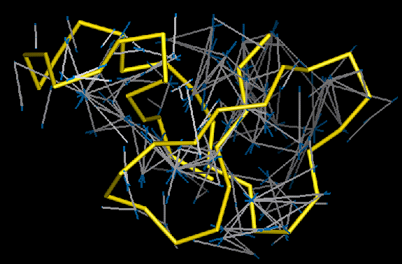
The figure above shows how the satisfied distance restraints output by viol2pdb are depicted in QUANTA.
The solid yellow bonds show the protein's C-alpha backbone trace. The white sticks, with their blue extensions, correspond to the satisfied NOE distance restraints. Each white line joins two restrained atoms while the blue extensions correspond to the overall length of the upper-bound restraints defined for those two atoms.